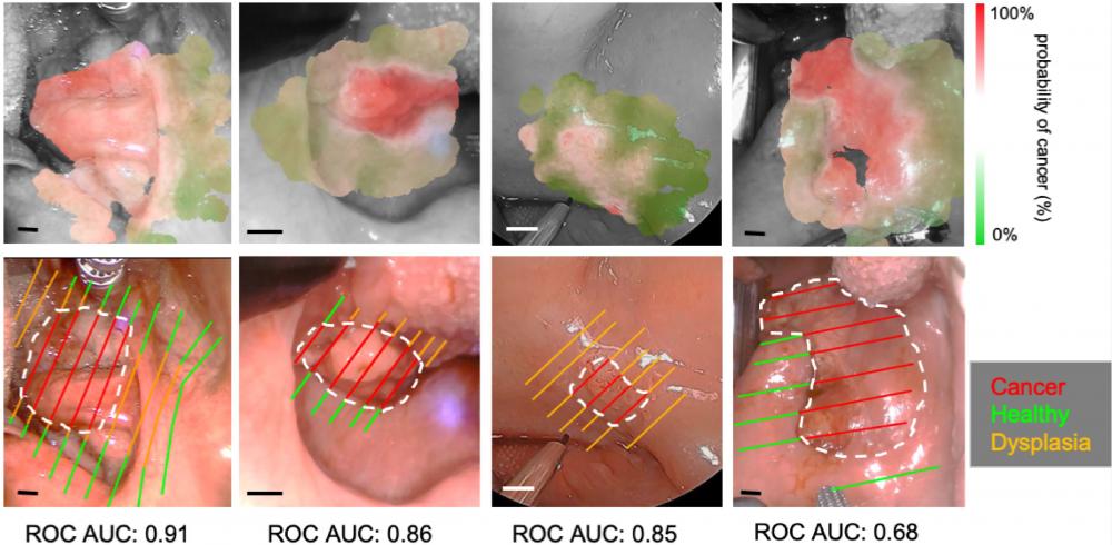
Classification visualizations and histological slice annotations for four in vivo patient scans (RF method used). Only labels derived directly from histology are shown with cancer outlined in white. Additional labels for homogeneous regions (healthy/cancer only) were also employed for model training. Clear deliniation of cancer regions is observed, even for patients with a lower AUC score. This is due partially to the presence of dysplasia which is omitted from training/validation as well as a small margin of error with histological coregistration. The scale bar corresponds to 5 mm in all cases.
This research involves the application of clinical Fluorescence Lifetime Imaging (FLIm) for intraoperative detection and surgical guidance of Head and Neck cancer. Cancers of the oral cavity (e.g., tongue, gingiva, floor of mouth) and oropharynx (base of tongue, palatine tonsils, lingual tonsils) were evaluated in a 100-patient research effort over a period of 2017 to 2021. A subsequent 100-patient research cohort is to be enrolled from the present (2021) onward to investigate further research aims.
Current progress has demonstrated that FLIm can be successfully integrated into existing surgical workflows, such as the da Vinci Transoral Robotic Surgical Platform. Collective results have demonstrated that FLIm can delineate healthy tissue, high-grade dysplasia, and cancer through both univariate analysis of time-resolved and intensity-based autofluorescence, as well as through machine learning classification approaches to enhance surgical decision-making.
Ongoing work seeks to account for the degree of inter-patient data heterogeneity arising from distinct cancer types, medical histories, patient demographics, and molecular features of tissue. New methods for augmenting FLIm data, implementing real-time classifiers, and accounting for surgical motion in real-time will continue to be investigated and refined in ongoing work.
Collaborators
Dr. Andrew Birkeland, M.D. (UC Health)
D. Gregory Farwell, M.D. (UC Health)
Dr. Marianne Abouyared, M.D. (UC Health)
Dr. Arnaud F. Bewley, M.D. (UC Health)
Dr. Dorina Gui, M.D., Ph.D. (UC Health)
Jonathan Sorger, Ph.D. (Intuitive Surgical)
Funding
NIH (National Institutes of Health): R01 CA187427
Publications
Weyers, B. W., Birkeland, A. C., Marsden, M. A., Tam, A., Bec, J., Frusciante, R. P., Gui, D., Bewley, A. F., Abouyared, M., Marcu, L., & Farwell, D. G. "Intraoperative delineation of p16+ oropharyngeal carcinoma of unknown primary origin with fluorescence lifetime imaging: Preliminary report." Head & neck, 44(8), 1765–1776 (2022).
M. Marsden, B. W. Weyers, J. Bec, T. Sun, R. F. Gandour-Edwards, A. C. Birkeland, M. Abouyared, A. F. Bewley, D. G. Farwell, and L. Marcu, "Intraoperative Margin Assessment in Oral and Oropharyngeal Cancer Using Label-Free Fluorescence Lifetime Imaging and Machine Learning," IEEE Trans Biomed Eng 68, 857-868 (2021).
Weyers BW, Marsden M, Birkeland AC, Frusciante RP, Bec J, Tam A, Sun T, Gui D, Bewley AF, Abouyared M, Marcu L, Farwell DG. Intraoperative Label-Free Fluorescence Lifetime Imaging for Real-Time Delineation of Oropharyngeal Carcinoma of Unknown Primary Origin. Oral Oncology. Under Peer Review (April 2021).
M. Marsden, T. Fukazawa, Y. C. Deng, B. W. Weyers, J. Bec, D. Gregory Farwell, and L. Marcu, "FLImBrush: dynamic visualization of intraoperative free-hand fiber-based fluorescence lifetime imaging," Biomed Opt Express 11, 5166-5180 (2020).
B. W. Weyers, M. Marsden, T. Sun, J. Bec, A. F. Bewley, R. F. Gandour-Edwards, M. G. Moore, D. G. Farwell, and L. Marcu, "Fluorescence lifetime imaging for intraoperative cancer delineation in transoral robotic surgery," Transl Biophotonics 1, e201900017 (2019).